27 diciembre 2022
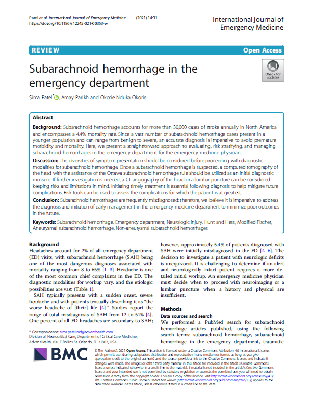
Manejo de la hemorragia subaracnoidea en los servicios de Urgencias
La hemorragia subaracnoidea es una entidad clínica relativamente frecuente (30.000 casos anuales en Estados Unidos) y con una mortalidad no desdeñable. Su presentación urgente, en el contexto de una cefalea, obliga a una cuidadosa aproximación a este signo para evaluar, estratificar el riesgo y manejar las hemorragias subaracnoideas en los Servicios de Urgencia Hospitalarios (SUH). Este artículo de revisión, publicado por tres intensivistas del hospital AdventHealth de Orlando (Florida) repasa los síntomas que la sugieren, las pruebas diagnósticas a realizar y su clasificación, y aporta unas pautas para su manejo tanto médico como quirúrgico. Si te interesa puedes descargarlo aquí: https://drive.google.com/file/d/1qVGFk0i2OEllc4Ge0o8R1H9rNMMkGA9d/view?usp=sharing
Subarachnoid hemorrhage is a clinical entity quite frequent (30,000 cases annually in North America) that encompasses a 4.4% mortality rate. Its typical presentation, in the context of a headache, is imperative an accurate diagnosis of this symptom. This article, published for three intensive care doctors from AdventHealth Hospital in Orlando (Florida, USA), present a straightforward approach to evaluating, risk stratifying and managing, medically and surgically, subarachnoid hemorrhages in the ED. Download link: https://drive.google.com/file/d/1qVGFk0i2OEllc4Ge0o8R1H9rNMMkGA9d/view?usp=sharing
14 diciembre 2022
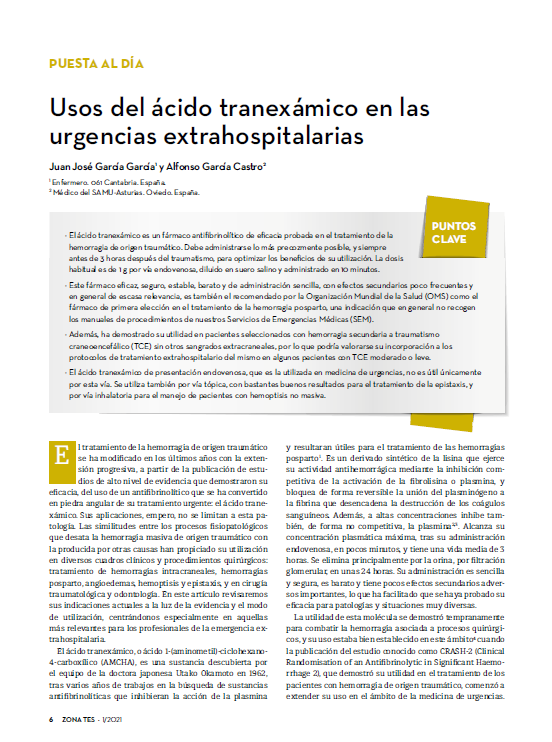
Usos del ácido tranexámico en las urgencias extrahospitalarias y la hemorragia postparto
El ácido tranexámico es un fármaco antifibrinolítico de eficacia probada utilizado para la hemorragia de origen traumático, administrado idealmente dentro de las tres horas siguientes al traumatismo por vía endovenosa (1 g diluido en 100 cc de suero salino a pasar en 10 min es la pauta más habitual). También ha demostrado su utilidad en pacientes seleccionados con hemorragia secundaria a traumatismo craneoencefálico sin otros sangrados extracraneales y en determinados pacientes con angioedema, y por vía tópica o nebulizada en pacientes con hemoptisis o epistaxis. Todos estos usos se revisan en el artículo de 2021 que puedes descargar en este enlace: https://drive.google.com/file/d/1reaVC0x87TozVFE08c5FvB7KCqiDkSip/view?usp=sharing
Ese artículo repasaba también su papel en el manejo de la
hemorragia postparto, concluyendo que, a la luz de la evidencia disponible en
aquel momento, era el de elección, por recomendación de la OMS-WHO, para su
administración en las tres primeras horas tras el parto. Una exhaustiva
revisión publicada en ese mismo año en la Revista Chilena de Anestesia confirma
la misma recomendación, y propone considerar su uso en pacientes con alto
riesgo de hemorragia que vayan a ser sometidas a una cesárea. Puedes
descargarlo en este enlace:
01 diciembre 2022
Fármacos y fluidos administrados por vía intraósea en pacientes críticos
19 noviembre 2022
El manejo con control de la temperatura en pacientes con parada cardiaca
https://drive.google.com/file/d/158mXv4R3WZzO4YJdEbHGDyYm6VYz3aPz/view?usp=sharing
Targeted temperature management (TTM) is a technique which
has been in use as a part of the post-cardiac arrest care for almost 20 years,
since the first major reports on its benefits in improving out-of-hospital and
in-hospital cardiac arrest survival as well as improving neurological outcome.
However, the findings from one of the biggest trials to date in this topic, the
TTM2 trial, surprisingly showed no difference in mortality between patients
after out-of-hospital cardiac arrest who underwent normo- or hypo-thermia. This
results, far from contraindicate its use, forces us to re-evaluate with new
perspectives this technique in order to discover why the demonstrated beneficial
effects in animal models aren’t replicated in humans. Download link:
04 noviembre 2022
Anatomía del sistema arterial coronario
If you're familiar with the anatomy of the coronary arterial system you can deduce quite precisely, when interpreting a 12-lead ECG, which will be the heart area affected by an acute coronary syndrome. The document attached to the download link reviews systematically the main branches of the coronary system, the territories that supply and the associated miocardyal infarction caused by its acute obstruction. Download link: https://drive.google.com/file/d/1YSDRFO4cxLQlUlT6bxw23sOQy-XeZ-IA/view?usp=sharing
19 octubre 2022
Manejo extrahospitalario avanzado de pacientes con hemorragia no compresible tras un traumatismo penetrante
04 octubre 2022
Eficacia de las maniobras de compresión abdominal para aumentar la presión intratorácica
The Heimlich manouevre is a well-known intervention for the management of choking due to foreign body airway occlusion, but the evidence base for guidance on this topic is limited. The authors of this article, researchers from the Imperial College of London, measured pressures during abdominal thrusts in healthy volunteers. Their interesting findings are quickly summarized in the abstract, but the article is not that long that you can read it all… Download link: https://drive.google.com/file/d/1_1l0CeUOcgEPDV0cM6AtJa2zkfTV7gX4/view?usp=sharing
29 septiembre 2022
Prevención de complicaciones en los accesos intravasculares
https://drive.google.com/file/d/1GifKM6yzh0_G-j8MSRw7k6SDdrvP6xdg/view?usp=sharing
Intravascular access is mandatory, in many scenarios, for prompting resuscitation, and eventually monitoring, our critical patients. Mechanical complications at insertion, and infections and thrombosis during catheter use are quite frequent, with a reported incidence of more than 60 episodes for 1000 catheter-days. Therefore, necessity is what should guide our catheter use, always preferring the device with the lowest complication rate or the less invasive one. This article reviews the principles that should guide the election of the intravascular access, and also the key prevention measures of catheter infections. Download link: https://drive.google.com/file/d/1GifKM6yzh0_G-j8MSRw7k6SDdrvP6xdg/view?usp=sharing
26 septiembre 2022
Algoritmo de Brugada: TV vs TSV con conducción aberrante
Este artículo, publicado en la revista Circulation en 1991, proponía, tras haberlo testado prospectivamente en 554 pacientes, un método en cuatro pasos consecutivos para discriminar, dentro de las taquicardias de complejo ancho, cuáles eran taquicardias ventriculares (TV) verdaderas y cuáles eran taquicardias de origen supraventricular (TSV) con conducción aberrante. Aunque los impresionantes niveles de sensibilidad (98,5%) y especificidad (96,7%) obtenidos en ese estudio han sido muy matizados por estudios posteriores, el algoritmo de Brugada sigue siendo una de las principales herramientas electrocardiográficas para discriminar entre TV y TSV con aberrancia. Enlace de descarga: https://drive.google.com/file/d/18IofGHbcBVx4G54zq85FUIF4IOdKvPn6/view?usp=sharing

This article, published in Circulation in 1991, proposed, after having tested it prospectively in 554 patients, a stepwise approach to do a differential diagnosis between ventricular tachycardia (VT) and supraventricular tachycardia (SVT) with aberrancy. Although the impressive levels of sensitivity (98.5%) and especificity (96.7%) reported by Brugada et al have been very shaded by other studies, Brugada criteria remain one of the main ECG criteria tools to discern between VT and SVT. Download link: https://drive.google.com/file/d/18IofGHbcBVx4G54zq85FUIF4IOdKvPn6/view?usp=sharing
23 septiembre 2022
Revisión de las principales asincronías paciente-ventilador
Patient-Ventilator Dyssynchrony (PVD) is often described as a patient “fighting” the ventilator. In fact, there are many forms of dyssynchrony some of which can very subtle. If unrecognized early, dyssynchrony can evoke patient discomfort, increase incidence of lung injury, lead to oversedation, and lengthen duration of mechanical ventilation.The figures presented in this article depict dyssynchrony in Volume Control Ventilation (VCV) with a decelerating ramp of flow, taking into account their causes and the corrective measures to take. You can download it here: https://drive.google.com/file/d/1l--Kwv_N-V27Uofu72-xrQ9AquoMBFGg/view?usp=sharing.
13 septiembre 2022
El retraso en la administración de adrenalina en pacientes con PC en AESP se asocia con peor pronóstico neurológico
El número de pacientes con buen estado neurológico tras superar una parada cardiaca con ritmo inicial desfibrilable ha ido aumentando en los últimos años como resultado de la progresiva expansión del uso de desfibriladores de acceso público. Sin embargo, no sucede lo mismo en los pacientes con ritmo inicial no desfibrilable, y además se observan diferencias entre los dos ritmos con estas características: mientras que va aumentando la supervivencia de pacientes con actividad eléctrica sin pulso (AESP) como ritmo inicial, la de los que debutan con asistolia permanece estancada. Esto lleva a algunos autores a proponer un tratamiento diferenciado para ambos ritmos. En este contexto, los autores de este artículo japonés han buscado establecer, en una revisión retrospectiva de 4.168 pacientes con parada cardiaca presenciada con AESP como ritmo inicial, si la demora en la administración de adrenalina puede influir en el pronóstico neurológico de estos pacientes. La conclusión del artículo es que sí: cada minuto de retraso en la administración de adrenalina se asocia con una disminución del 6% de las posibilidades de sobrevivir con buen estado neurológico (CPC 1-2 a los 30 días), y las posibilidades disminuyen rápidamente cuando la demora supera los 10 minutos. Puedes descargarte este interesante artículo en el siguiente enlace: https://drive.google.com/file/d/1yjVTscajLv66DV5K3QD7XemDk2S1Z5tU/view?usp=sharing
The number of survivors with good neurological status in patients who suffered a cardiac arrest with an initial shockable rhythm has been increasing in the last years as a result of the rise of the use of public access defibrillators. However, there isn’t a similar trend in patients with an initial non-shockable rhythm. There are also differences between them: survival in patients with pulseless electrical activity (PEA) as initial rhythm is increasing, while not in patients with asystole recorded as initial rhythm. Some authors propose to treat differently both rhytms, and is in that context in which the authors of this study aimed to determine, in a retrospective review of 4.168 patients with witnessed OHCA with PEA as initial rhythm who received epinephrine, whether the time to the administration of that drug could affect the neurological outcomes in patients with these characteristics. And they conclude that it certainly affects: each minute of delay in the administration of epinephrine is associated with a 6% reduction in the likelihood of achieving favorable neurological outcome (CPC 1-2 at 30 days), and the odds of it rapidly decreased after 10 min. You can download this interesting article in the following link: https://drive.google.com/file/d/1yjVTscajLv66DV5K3QD7XemDk2S1Z5tU/view?usp=sharing
07 septiembre 2022
El origen del test de Apgar
En 1953 la anestesista obstétrica Virginia Apgar publicó un artículo en el que daba a conocer los resultados de un estudio para establecer unos criterios para establecer, de forma sencilla y clara, la necesidad de reanimación de un recién nacido en un intento de disminuir la mortalidad infantil en las primeras 24 horas de vida. Para elaborar la escala, que hoy lleva su nombre y es de uso universal, buscó cinco parámetros que pudiesen ser evaluados de forma sencilla con la menor carga de subjetividad posible. Lo consiguió, como reconoce en el artículo, en cuatro de ellos, porque la evaluación de la cianosis fue "con mucho el menos satisfactorio de los signos", cuya evaluación "fue el motivo de las mayores discusiones entre los observadores" por una serie de factores que enumera en el artículo, todavía interesante de leer hoy en día en su primera parte (la que describe los parámetros). Fue republicado en 2015 por la Society of Obstetric Anesthesia and Perinatology, y lo tienes disponible en este enlace por si te apetece echarle un vistazo: https://drive.google.com/file/d/1ttAxboUODiV1ZhsU_2HRGSdCST2bXfWa/view?usp=sharing.
In 1953 the obstetric anesthesiologist Virginia Apgar issued an article to show the world the results of a study aimed to establish some criteria to determine, in a clear a simple way, the need of resuscitation in a newborn child. She looked for five items relevant for that purpose that could be determined with low variability among the observers to create a score. She got it with four of them, because the color (the presence or absence of cyanosis) was, in her own words, "by far the most unsatisfactory sing and caused the most discussion among the observers". Despite this, the score that bears her name in honor is nowadays an universal tool to determine the need of resuscitation in the newborn. If you'd like to take a look at its foundations we can do it following this link thanks to the Society of Obstetric Anesthesia and Perinatology, that republished the article in a digital format: https://drive.google.com/file/d/1ttAxboUODiV1ZhsU_2HRGSdCST2bXfWa/view?usp=sharing.
05 septiembre 2022
Urgencias en paciente portador de DAI
Un cardiólogo del Hospital Vírgen de las Nieves de Granada (España) publicó hace ya unos años, en la Revista Española de Cardiología, un interesante artículo de revisión de las urgencias que presentan, en relación con el dispositivo, los pacientes portadores de un desfibrilador automático implantable. En él aborda, primero, cuestiones generales sobre el manejo de estos pacientes: cómo ha de hacerse una evaluación general de los mismos, cómo influye su presencia en el manejo de la parada cardiaca o en la realización de técnicas diagnósticas... A continuación repasa las urgencias, tanto arrítmicas (descargas múltiples, síncope) como no arrítmicas (infección, trombosis, insuficiencia cardiaca congestiva), abordando tanto su forma de presentación como su manejo urgente.
Si te interesa puedes descargarlo en el siguiente enlace: https://drive.google.com/file/d/18VH7lx8ZRGZ4YSUvBbwnV27PCDSGypsY/view?usp=sharing

.png)

.png)


.png)


.png)


.png)

